Most people know they should get their moles checked. But if you’ve never actually done it, you probably have no idea what to expect when you walk into that clinic. Is it uncomfortable? Will they shave something off? Do they look at every single mole on your body?
These are fair questions and the answers are simpler than you’d think. Here’s a clear, honest look at what a professional skin check involves, why it matters, and what the doctor is actually looking for when they examine your skin.
Why Skin Checks Are More Important Than Most People Realise
Skin cancer is the most common cancer in Australia. According to the Australian Institute of Health and Welfare, two in three Australians will be diagnosed with some form of skin cancer by the time they turn 70. That’s not a statistic to brush off.
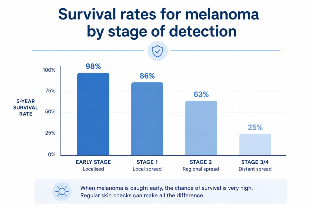
The good news is that when it’s caught early, most types of skin cancer including melanoma, the most dangerous kinds are highly treatable. The five-year survival rate for melanoma detected at an early stage is over 98%. Once it spreads, that number drops sharply.
This is why regular skin checks aren’t just recommended. For many people especially those with fair skin, lots of moles, or a history of sun exposure they can be lifesaving.
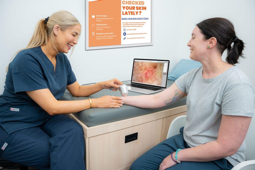
What Actually Happens When You Go for a Skin Check
When you arrive, the doctor, usually a GP or dermatologist will ask you a few questions. They’ll want to know your skin type, whether you’ve had significant sun exposure over your life, if you’ve ever had any skin cancers removed, and whether anyone in your family has had melanoma.
Then comes the physical examination. The doctor will ask you to undress (you’ll have a gown) so they can look at the skin across your whole body including your scalp, between your toes, and under your nails. These are spots that people miss when checking themselves.
The doctor looks for anything that stands out. Moles are assessed using a simple rule called the ABCDE criteria:
- A – Asymmetry: one half of the mole doesn’t match the other
- B – Border: edges that are irregular, ragged, or blurred
- C – Colour: multiple shades or uneven colour within one mole
- D – Diameter: anything larger than 6mm (about the size of a pencil eraser)
- E – Evolution: any mole that is changing in size, shape, or colour
If something looks concerning, the doctor will examine it more closely using a tool called a dermatoscope, a handheld magnifying device with a light. It lets them see the layers beneath the surface of the skin that the naked eye can’t detect.
Mole Mapping: The Next Step Up in Skin Cancer Screening
For people with many moles, a history of skin cancer, or a high-risk profile, doctors often recommend mole mapping. This is a more thorough process where high-resolution photographs are taken of your entire body and each individual mole.
The images are stored digitally, and at your next appointment, the doctor compares them side by side to detect even tiny changes that might not be obvious during a standard check.
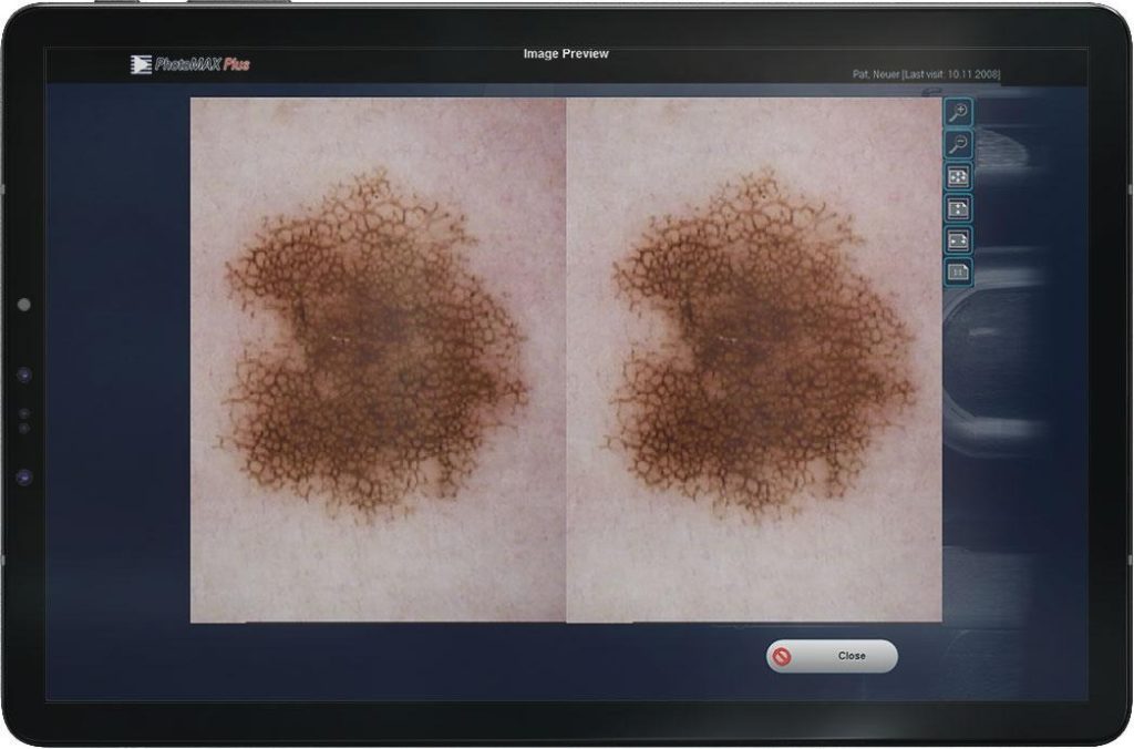
The technology used in clinics for this purpose has come a long way. Clinics that offer mole mapping typically use a purpose-built
Clinics that offer mole mapping typically use a purpose-built digital skin imaging system that captures standardised, high-resolution images of every lesion. This allows for precise comparison over time, something that’s simply not possible with the naked eye or a basic camera.
What Happens If the Doctor Finds Something Suspicious
Finding something suspicious during a skin check doesn’t automatically mean it’s cancer. In many cases, the doctor will simply note it, photograph it, and ask you to come back in three to six months so they can monitor it.
If they’re concerned enough to act immediately, they may perform a biopsy. This is a quick, in-clinic procedure where a small sample of the mole is removed under local anaesthetic and sent to a pathologist. It’s usually painless and takes less than 10 minutes. Results come back within a couple of weeks.
If the result shows skin cancer, the next steps depend on the type and stage. Basal cell carcinomas and squamous cell carcinomas the more common and less aggressive types are usually removed surgically during a simple clinic procedure. Melanomas may require a wider excision and further testing.
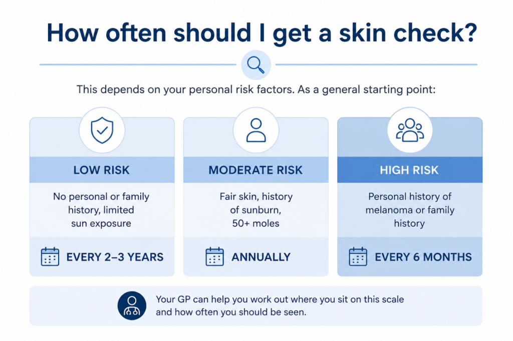
How Often Should You Get a Skin Check?
This depends on your personal risk factors. As a general starting point:
- Low risk (no personal or family history, limited sun exposure): every 2 to 3 years
- Moderate risk (fair skin, history of sunburn, 50+ moles): annually
- High risk (personal history of melanoma or family history): every 6 months
Your GP can help you work out where you sit on this scale and how often you should be seen.
A Few Things Worth Knowing Before You Go
Don’t paint your nails before the appointment. Nail beds can sometimes show early signs of melanoma, and nail polish makes this harder to see.
Wear your hair out or bring a hair tie. The doctor will want to check your scalp, and this is easier with your hair down.
Avoid wearing heavy makeup if you can. Some facial lesions can hide under coverage.
And bring a list of any moles you’ve noticed changing. Even if you’re not sure it’s significant, it’s worth mentioning.
The Bottom Line
A skin check is one of the most straightforward, low-risk things you can do for your long-term health. It takes 20 to 30 minutes, it’s not painful, and if something is caught early, it could make an enormous difference.
If you haven’t had one recently or ever book one. Your future self will thank you.
| Did You Know? |
|---|
| Modern mole mapping devices can capture and compare hundreds of skin lesions across multiple visits, giving doctors a level of precision that was impossible just 15 years ago. Ask your skin clinic whether they use digital imaging for long-term monitoring. |